Contrast to EEG
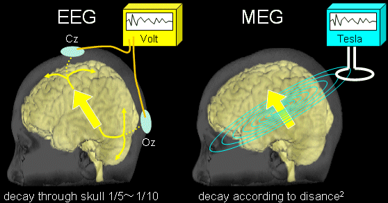
Magneto-Encephalo-Graphy detects electric brain activities as well as Electro-Encephalo-Graphy.
While the EEG detects the electric potential difference between two electrodes,
MEG detects the dynamic magnetic fields derived from their electric activites.

The electric signals through the skull are blurred and attenuated to a fifth - tenth level,
however, the magnetic signals through the skull are not.
With this reason, MEG can detect more sharp brain signals than scalp EEG.
The MEG signals are attenuated
- according to the second power of the distance between the current and the detector.
- if the current direction is nearly normal to the detector plane.
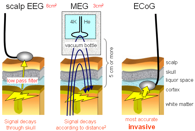
The sensitivity of scalp EEG, MEG, and Electro-Cortico-Graphy (ECoG) is
scalp EEG < MEG < ECoG
in ascending order.
To detect electric cortical signals, the scalp EEG requires wider than 6cm2 synchronized cortical activation,
while MEG requires wider than 3cm2.
The ECoG is the most sensitive, however, ECoG requires "craniotomy".

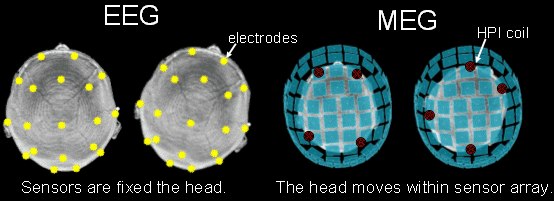
EEG electrodes are fixed to subjects' scalp and
the positional relationship between electrodes and the head remains unchanged
even if subjects move their heads during EEG examination, but not MEG examination.
MEG sensors are not fixed to subjects' scalp,
while three or more Head Position Indicator coils (HPI coils) are fixed to subjects' scalp in MEG.
Precedently, electricity is carried to HPI coils and HPI positionss on MEG coordination are calculated
(by means of electrically equivalent current dipole estimation).
Once the positional relationship between MEG sensors and HPI representing scalp position is established,
subjects' MEG data are recorded.
For the above reason, if subjects move their heads, the established positional relationship is lost.
HPI coil positions must be remeausred and MEG data should be recorded again.
The subjects are required to be immobile during MEG examination.


